In this article researchers report on experiments to improve understanding of the basic physical properties of imaging fiber bundles for applications in coherence imaging such as optical coherence tomography.
Using an imaging spectrograph with a large, sensitive detector on the end of an optical interferometer allows the team to perform detailed measurements of fiber bundles. Their results show that current commercial fiber bundles are not yet suitable of coherence imaging applications and encourage further research and development in that area.
Understanding the properties of DNA in different physical and chemical environments is important for biomedical and life science applications.
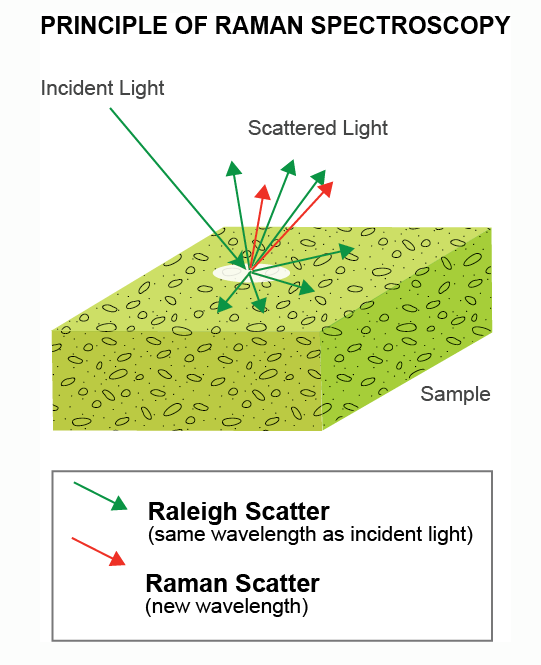
Sensitive, high resolution Raman spectroscopy gives information about the structure of DNA molecules. Often the signals are/need to be enhanced by processes like surface enhanced Raman spectroscopy to obtain measurable signals more effectively. Here, researchers measure the structural changes of DNA inside photonic crystal cavities.
Researchers around James Rice from Dublin, Ireland, are using self-assembled, highly ordered structures of gold nanoparticles to demonstrate molecule sensing at ultralow concentrations and excitation powers.
The team uses surface enhanced Raman scattering (SERS) in the presence of the Au nanoparticles and perform microscopic Raman spectroscopy and imaging.
US researchers Amber Moody and Bhavya Sharma are working on sensitive bio sensing for early disease detection and better understanding of biological processes. Here they show detection of neurotransmitters like melatonin, dopamine and others.
They use surface enhanced Raman spectroscopy (SERS) where the Raman signal is enhanced using metal nanoparticles. The Raman spectrum gives a clear fingerprint spectral signature. The researchers systematically study the effects of Au and Ag nanoparticles as well as different excitation wavelength and perform PCA analysis to establish the best conditions to achieve the lowest possible limits of detection. Sensitive spectroscopy systems provide resolution and sensitivity necessary for doing their measurements.
Researchers around Anja Royne and Joanna Dziadkowiec from Norway recently published an article about their experiments investigating surfaces of calcites. This mineral has multiple applications as a building and industrial material, and it is often important to characterize the strength of the materials involved. For example, depending of the surrounding medium (could be water) the formation of cracks and weakening of the material structure can be induced.
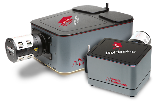
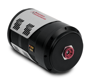
In their experiments they use rough and reactive calcite surfaces and perform measurements in a device for surface force measurements. The device uses an interferometric readout using a high-resolution imaging spectrometer (Isoplane 320, PIXIS 2048B). The interference effect allows for very precise measurements of the distance between the sample surfaces.
Silica based materials that emit light under excitation have huge applications in creating novel sensors, laser sources and optical devices in general. Silica can be doped with different elements to tailor its emission properties for different applications, however the interactions and exact mechanisms determining the emission are often complex and not easy to understand. So various techniques are used for characterization of these materials.
Researchers around Prof. Ouerdane from Univeristy Lyon in France are probing microstructured silica fibers doped with Bi ions using various forms of photoluminescence spectroscopy. The PL emission changes when the fibers are exposed to gamma rays, with laser excitation energy and with time after a laser excitation pulse. The results give the researchers new insights into the structure of the Bi color centers in the silica fibers.
The team performed the time resolved PL and PL excitation measurements using a spectroscopy setup and a PI-MAX4 ICCD camera as detector for experiments with high sensitivity and time resolution from nanoseconds to microseconds.
The use of catalysts in the chemical industry is common in the production process. However how these catalysts work exactly is not always known. The research group of Prof. Prashant Jain is using surface enhanced micro Raman spectroscopy (SERS) to examine the exact mechanisms of an Ag nano particle catalyst for chemical formation of ethylene.
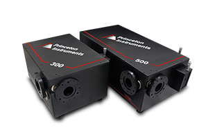
SERS gives the researchers a detailed look what chemical processes happen during the catalytic reaction. In the experiment a single Ag nanoparticle is observed spectroscopically. A 300m focal length SpectraPro spectrometer with a Pylon camera record the Raman spectra. The SERS observations show that the formation of graphene happens as a first step during the chemical process. Knowledge of the precise reaction steps then allowed the team to design a new combined graphene/Ag nanoparticle catalyst which enables the chemical process under ambient conditions using photoexcitation whereas the typical reaction scheme requires cost intensive high pressure and high temperature environments.
The team hopes to that their newly designed reaction will be used to optimize other catalytic reactions commonly used.
Optical spectroscopy plays a significant role in material science and researching properties of 2D materials. Raman and second harmonic generation spectroscopy shed light on the structure of materials and require sensitive detection using scientific spectroscopy systems.
2D materials are a class of materials that can be produced in atomically thin, crystalline layers (down to single atomic layer thickness). Graphene, transition metal dichalcogenides and other 2D materials have shown to have unique physical properties that make them appealing for development of new, enhanced electrical and optoelectronic devices. Over the past decade researchers have also started to build layered structures of 2D materials (sometimes referred to as vander Waals heterostructures) to build new devices for studying new physical effects or practical applications. Detailed knowledge of the individual materials is necessary to consider their use for new, interesting applications.
Researchers from Vilnius University (Lithuania) and Oak Ridge National Laboratory (USA) are studying a class of materials called thiophosphates for their ferroelectric properties and utilization of these properties in 2D vander-Waals heterostructures. Ferroelectric materials in analogy to ferromagnetic materials can possess an oriented, permanent electric polarization that can be changed with an electric field. The ferroelectric behavior is temperature dependent (with temperature being below a transition temperature for order to occur) and for thiophosphates it can also be studied as a function of thickness, where the material might prefer ferroelectric order or antiferroelectric order.
There also can be nanoscale ferro and antiferroelectric domains coexisting. The researchers are interested in understanding exactly the properties of thiophosphates on the nanoscale to “help to identify the possible mechanisms by which these materials can be functional in van der Waals heterostructures”.
The team uses a piezo response force microscopy (similar to AFM, but sensitive to electric polarization) to characterize thiophosphates, but the observations are supported by sensitive Raman spectroscopy and second harmonic generation spectroscopy as a function of temperature. Optical spectroscopy has always been one of the main characterization tools to characterize the structure of 2D materials and interactions of electrons and atoms in their crystal lattice. Both Raman spectroscopy and second harmonic generation spectroscopy are sensitive to the structure of materials. Raman measures the inelastic scattering from vibrational modes of the crystal lattice, while existence of SHG indicates a breaking of inversion symmetry which would imply some form of ordering in the probed volume for ferroelectric materials.
For Raman spectroscopy the researchers use a 532nm laser for excitation which is being kept in at a low power of 100µW. It is often important to work with low laser powers when studying materials to avoid effects from heating the sample or risking damage. However, signal levels will be low requiring sensitive detection of the Raman scattered light. For SHG a 50fs at 800nm and 80MHz rep rate with 0.1W of energy at the sample is used for excitation. In both cases appropriate filters block scattered light at the excitation wavelength.
In both situations a SpectraPro spectrograph with 300mm focal length and a PIXIS camera are used for spectroscopic detection. The researchers use the ability of this system to adapt to various experimental conditions. The spectrograph turret holds several gratings to optimize resolution and grating efficiency for either measurement. The scientific CCD camera is suitable for detecting ultra-low light level signals with high sensitivity and/or signals that require a high dynamic range, so optimal conditions for Raman and SHG measurements can be achieved.
Researchers at DOE (Department of Energy, United States) undertook experiments to compare temperatures measured simultaneously by pyrometry and Raman spectroscopy in shock-wave-compressed toluene. Comparisons of this type are necessary to validate pyrometry as a temperature diagnostic in dynamic experiments. Additionally, this is also helpful for opaque samples where pyrometric signal is contaminated with possible nonthermal sources of light emission, such as window fractoluminescence.
Temperature dependence of the intensity ratios of anti-Stokes to Stokes Ramanscattered lines of various vibration modes are utilized for the purpose of temperature calculations of the sample. 12 ns pulse length of excitation laser was used to excite the sample and Raman-scattered signal from sample was imaged onto a 300mm long by 1.5mm core quartz optical fiber. After passing through a filter assembly the light entered a fiber-optic bundle that formed a line on its output end at the entrance to an IsoPlane 160 spectrometer paired with an intensified charge-coupled device (ICCD) detector (PI-MAX4). ICCD was useful obtaining desired gain and gating around 12ns laser pulse.