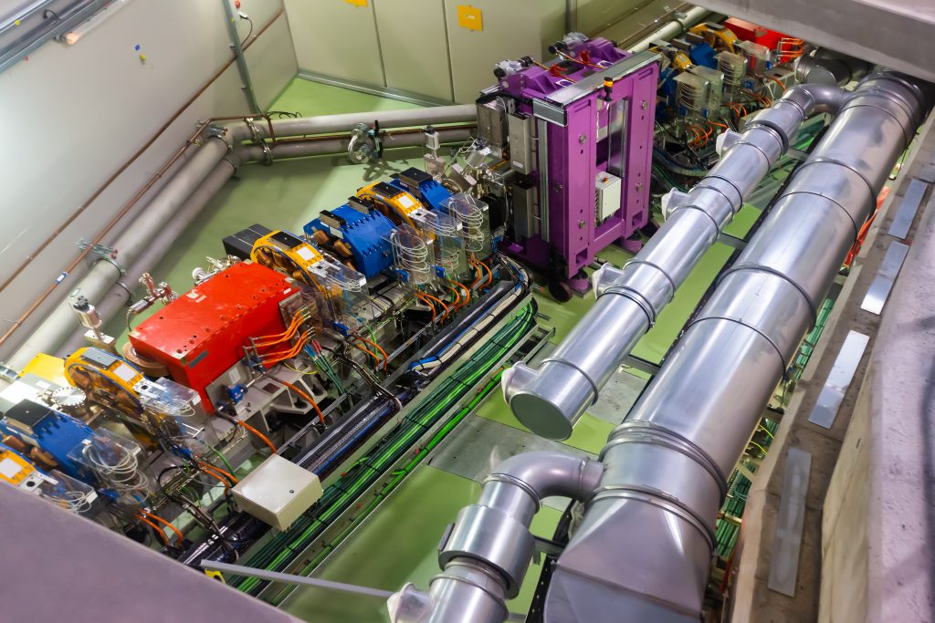
Scientists at the Institute for Pulsed Power Science of Kumamoto University in Japan are studying streamer discharge where a voltage difference is applied across an insulating gas or air. Streamers have a filamentous structure (think of the fine discharge arc from a tesla coil that are used for electricity shows and demos) and are a form of non-thermal plasma where the electron temperature is much higher than the ion temperature. Roughly speaking a fast-pulsed high voltage will mainly move atomic electrons whereas the atomic nuclei are unaffected due to their higher mass. Non thermal plasmas have found use in both ozone production and plasma medicine. Air purification has become an important application for non-thermal plasmas, as they can decompose mold, bacteria, viruses and other hazardous substances and pollutants such as NOx, SOx and volatile organic compounds. The group from Japan is currently most interested in ultrashort discharges with durations of about 5ns that turn out to be more efficient in air production. There also seems to be differences in efficiency between positive and negative streamers (relating to the change in polarity of the high voltage potential used in their production). The mechanisms for this efficiency increase are not yet well understood though.
The researchers recently published a conference paper where they describe how they use ultrafast imaging of the generated streamers on the nanosecond timescale to characterize their size as well as propagation distance and velocity. A new experimental system based on intensified CCD cameras (ICCDs) can take snapshots of the plasma discharge. ICCDs use an image intensifier tube that allows to apply a variable electronic shutter, called the gate, with opening times (gatewidth) down to sub nanosecond timescales and amplifies the signal using a variable gain so even low numbers of photons can be observed. The interframe time in ICCDs is much longer (100 times or more) than the lowest gatewidth. However, observing the dynamics of the sub 10ns plasma discharge requires acquisition of more than one snapshot image.
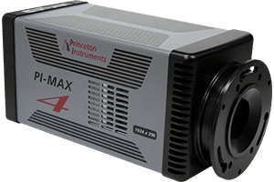
The researchers present a new experimental setup using a framing system based on 4 electron-multiplied ICCD cameras (PI-MAX4:1024EMB emICCDs). The cameras are setup to precisely trigger acquisition with 2.5ns delay between cameras so a sequence of 4 images taken within 10ns can be used to analyze the dynamics of the generated streamers. This corresponds to a burst frame rate of 400MHz. Besides the ultrafast timing, emICCD cameras are designed using a double gain mechanism, combining intensifier gain with on-chip signal gain which allows for stronger amplification of low light signals using ultrashort gatewidths. The double gain of the emICCD solves the problem of non-linear signal response of standard ICCDs. The combined gain of the camera allows for linear response over a wide intensity range, so low and high light intensity regions can be quantitatively analyzed.
This research shows that sensitive detection with multiple emICCD cameras is an effective way to analyze the dynamics of ultrafast phenomena and works well to characterize non-thermal transient plasma discharges on the nanosecond timescale.
New ceramic materials play and important role when higher temperature stability or performance in batteries is required. Researchers from Darmstadt (Gemany) are investigating ceramics based on carbon materials. The behavior of the material is strongly influenced by the phase and crystal structure of the carbon materials which can be probed by Raman spectroscopy.
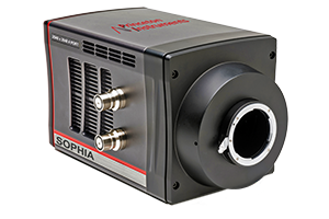
Specifically, the researchers are using UV Raman spectroscopy (excitation at 256.7nm) as it leads to stronger excitation of signals from some carbon structures. They use a high performance, multi-stage spectrograph as it can provide high resolution and filter capabilities with high stray light rejection at this wavelength (the range 0-1700 wavenumber is within a few nm of the excitation wavelength).
Researchers from Frank Reidy Research center for Bioelectric (Old Dominion University) have been investigating relatively short duration (>200ns) pulsed helium atmospheric-pressure nanosecond pulsed jets (APNPJs) in air using Thomson scattering (TS), measuring the electron properties of the jets.
They utilize similar APNPJ geometry and pulse parameters that have previously been applied for dental and biomedical applications. Their measurements have made an attempt to elucidate the role of electronic collisions in production of reactive plasma species and provide additional experimental validations for numerical models. For performing optical emission spectroscopy their setup uses a TriVista 555 triple grating spectrograph coupled to a PIMAX ICCD (GenIII) camera. The scattering images were rotated 90 degrees before being projected onto entrance slit of TriVista using pair of dichroic laser mirrors and spectral resolution of 0.17 nm was obtained using this detection system.
The next generation of high resolution EUV lithography require tools for reliable characterization and defect inspection of lithography masks.
Ideally the inspection is used with the same type of radiation and wavelength used in the lithography process. Researchers from the PSI in Switzerland demonstrate use of coherent, monchromatic EUV beams at 13.5nm to perform coherent diffraction imaging on lithography masks for defect inspection. New, more advanced image processing algorithms are applied to the image data taken with a large size, in vacuum CCD camera.
X-ray crystallography is an important tool for structure determination in material and biological science. Bright X-Ray sources like the Free electron laser at DESY in Hamburg can deliver intense X-Ray beams on very short timescales that allow probing of crystal structure on very small sample sizes.
Due to the nature of the X-Ray sources researchers require new means of sample delivery to the probe region. Here researchers developed means to deliver liquid streams of protein crystals to the interaction region while observing X-Ray diffraction as well as high speed optical imaging. In vacuum CCD cameras are used to collect the diffraction patterns.
Intense laser pulses make it possible to create high quality X-Ray radiation sources in a laboratory setting where typically large synchrotron sources are needed.
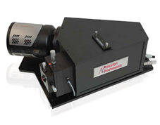
A research team from France has created such a source for ultrafast X-Ray absorption experiments on the femtosecond timescale. The researchers study what happens in the first few moments when a material is heated with a short, intense laser beam. The setup includes an X-Ray spectrometer where the radiation is detected with an in vacuum MTE camera.
Research facilities for X-Ray free electron lasers (XFEL) exist all around the world, having been developed over the past decade. The technology behind XFELs allows for the production of brighter and higher quality high energy radiation from EUV/Soft X-rays to hard X-rays. New and improved forms of fundamental physics measurements, from material science to structural biology, will be possible at these facilities.
The PAL-XFEL is a facility in Korea that opened for users in 2017. In addition to bright X-rays the facility provides pulsed beams with a small width below 50fs. This allows the measurement of dynamic processes, related to atoms and electronics in materials, at energies not available with other technologies. The facility operates several beamlines for hard and soft X-Ray experiments. Among other detectors a PI-MTE is used on the soft X-ray scattering and spectroscopy beamline, using the large 2048x2048px sensor size with the flexibility of mounting the detector in vacuum.
The PI-MTE is used in coherent X-Ray diffraction imaging as well as X-ray absorption spectroscopy experiments. In this article researchers from the facility describe measurements that characterize the performance of the instruments at the beamline. For example, the researchers measure magnetic circular dichroism in the absorption spectra of Co/ Pt multilayers using circular polarized X-Rays. These measurements allow researchers to make conclusions about the bulk ferromagnetism of this material.
Magnetic thin film materials play an important role for technological developments in sensing and memory elements. Using X-Ray scattering the structure and dynamic behavior of the magnetic films can be studied to understand their behavior in more detail.
Thin ferromagnetic films show interesting physical behavior and are considered to be of great interest for future sensing and memory applications. In a recently published report an international team of researchers in France, Italy, Slovenia and the UK report on some of their efforts to better understand the microscopic magnetic properties of thin film ferromagnetic materials and sandwich structures of ferromagnets and non-magnetic materials. These materials can show complex behavior such as formation of magnetic chiral structures and skyrmion lattice phases that are important for their physical behavior. The researchers were in particular interested in the dynamic formation of magnetic domains showing these complex structures.
To measure the magnetic characteristics of magnetic thin films one can use magnetic circular dichroism, where left and right circular light beams show different scattering behavior depending on the magnetization of the material. The researchers use magnetic dichroism to measure time resolved X-Ray magnetic scattering at the DiProI beamline of the FERMI free-electron laser in Trieste, Italy. The measurements utilize a pump probe measurement scheme where they induce demagntization in the sample material using a short 100fs laser pulse at 780nm, followed by a 60fs X-Ray probe beam at 20nm wavelength. This arrangement is sensitive to the magnetic rearrangement after the hit with the NIR pulse on picosecond timescales.
The X-ray reflected x-ray diffraction pattern is recorded by imaging with an in vacuum CCD (Princeton Instruments MTE 2048) that sits in close vicinity to the sample (12cm distance) to cover a large angular region of diffracted radiation. Imaging the different diffraction patterns of left and right circular polarized X-Rays can be used to determine the domain wall width and their dynamic behavior from the image data.
Conclusively the technique used by the research team was successful in detecting magnetization and domain properties in thin film ferromagnetic materials on picosecond timescale.
While the cutting-edge performance in astronomical observations is achieved by space based telescopes and ultra large telescope facilities on earth, researchers use and develop new smaller measurement facilities for specific measurement tasks and other applications that do not require the immense resources of larger facilities.
The Deep South Telescope (DST) is a 1m telescope facility (using a PW1000 telescope form PlaneWave instruments) that was built and is operated by the US Naval observatory (USNO) at the Cerro Tololo Interamerican Observatory in Chile, for observation of objects in the southern hemisphere. One of the measurements projects performed at the DST involves follow up observations confirming results of sky surveys performed by the Gaia satellite. The success of the Gaia space mission of the European Space Agency effectively ended ground based measurements, according to Norbert Zacharias and his coworkers at USNO. Gaia was launched in 2013 and has since catalogued the position and motions of billions of galactic objects with unprecedented measurement precision.
The DST was designed and optimized for astrometry, that is the precise measurements of the positions and movements of objects, stars and galaxies in the sky. In a recent paper, where the team around Dr. Zacharias reports on the installation and first measurements of the DST, they describe their goal to focus on smaller regions in the southern hemisphere to investigate and follow up on peculiar results from the Gaia measurements in collaboration with the Paris Observatory. Specifically, researchers noticed that the position of around 10% of objects in the international celestial reference frame (ICRF), obtained via ultra-precise, long-baseline radio-interferometry, deviate significantly from the positions obtained by Gaia taken at optical wavelength of the electromagnetic spectrum. Understanding the differences of these measurements is important as the ICRF is the standard reference frame for definition of the position of astronomic objects.
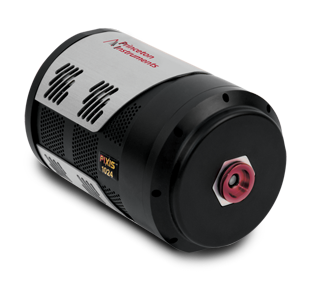
For optical detection at the DST it was important to the researchers to have a highly sensitive detector that covers a large amount of the focal plane of the instruments, alongside offering a high dynamic range, to observe faint objects as well as bright stars. The telescope uses a Teledyne Princeton Instruments SOPHIA camera with a large 4k x 4k sensor to achieve their measurement targets. Based on their first light tests they note that “the dynamic range of the instrument is very large, about 8 magnitudes, due to the relatively large pixels, sampling and low noise properties of the detector running at -80⁰C cooled by TEC with glycol (no liquid nitrogen, no dewar).”
Currently the researches are troubleshooting small issues, that occur at every new telescope facility upon first light, and they hope to obtain first measurement results during the year 2020. However, these results show that the new generation of commercial, larger sensor area CCD cameras are a suitable choice for challenging astronomical observations.
Cancerous tissue can be identified using Raman spectroscopy. In this paper researchers use Raman spectroscopy, reflectance spectroscopy as well as white light and autofluorescence imaging combined to identify cancer tissue with high fidelity.