Chemiluminescence Imaging in combustion experiments helps identify the size and shape of the reaction region when fuel is burned.
Most important is the emission from OH and CH radicals at 307nm and 430nm. An ICCD with short gating and using appropriate filters can takes snapshots of the luminescence of either molecule.
In the last few years the group of Brian Pogue has used ICCD cameras to image Cherenkov light emitted from patients’ bodies during radiation therapy with high energetic particles/photons.
The benefit is that the patient can be kept in room light which is suppressed by using small gate times only collecting light during short radiation pulses. Here they show how similar techniques are used for surface dosimetry, where they look at scintillator targets simultaneously to the Cherenkov light emission. The technique delivers fast and accurate calculation of the delivered energy dose to the patients.
Researchers around Mingxing Jin are studying luminescent effects in plasmas that are generated by intense laser pulses going through a gas.
Their goal is to identify processes that can maximize the luminescent lifetime in the gas. In their setup they shine a laser beam through a fast-moving gas jet and detect the spatial patterns of luminescence by taking quick snapshots at different times after the laser pulse.
Spectroscopic analysis of the signal is used to identify the nature of the light emitting molecules. The experiments demonstrate well the practical applications of an ICCD camera for doing fast, time resolved measurements on the nanosecond to microsecond timescale.
Raman spectroscopy plays an important role in assessing properties of tissue used in vascular reproductive surgery. Its power is extended by correlation with other optical measurement techniques such as FLIM and correlating the observations. Fundamental studies identify indicators in the Raman spectra to quantify structural or biochemical changes in tissue to monitor its quality before transplantation and over time.
The application of Raman spectroscopy in life science and specifically medical diagnostics seeks to utilize its advantages over traditional or established measurement techniques. Raman spectroscopy can identify biochemical substances with high specificity, revealing lots of details about tissue. It is non-invasive and requires no or only little sample preparation. Measurement results are output quickly with potential diagnostic results within seconds or minutes. Moreover, as Raman spectroscopy is an optical measurement technique, it can be easily combined with other optical measurement techniques enabling potential for correlation to achieve better diagnostic results. The optical integration for microscopy or in vivo measurements can be easily achieved using fiber optic systems.
As the biochemistry and structure of tissue is complex so are the Raman spectra collected from it. For every diagnostic application it is important to demonstrate how effective and accurate the results from the optical measurement technique are. In this process the specific molecular changes that indicate targeted change in tissue must be identified, that is specific changes in intensity and wavelength of spectral lines in the Raman spectrum. Then these changes can be targeted using advanced analysis to obtain quantitative diagnostic results.
Researchers around Prof. Juergen Popp from Friedrich Schiller University in Jena (Germany) and Prof. Laura Marcu from UC Davis (USA) performed experiments to characterize and diagnose molecular changes in bovine pericardium (BP) using optical measurements. Their optical approach combines Raman spectroscopy and fluorescence lifetime imaging (FLIM).
BP is a biomaterial used in reconstructive surgery, particularly related to heart valve replacements and vascular bypasses. The main failure mode of vascular transplants is calcification degrading the mechanical stability of the implant as well as increasing the potential for life threatening blood clots and thrombosis. As BP is derived from animal tissue it needs to be specially prepared and chemically stripped from its potential to induce a negative immune response in the host body. After this process BP needs to be mechanically stabilized by chemically crosslinking the collagen network building the tissue. However increased crosslinking has been attributed with increased risk of calcifications. The scientists note in their article that “there is a need for biochemical evaluation of the tissue properties prior to implantation to ensure that quality and reliability standards are met”. So, a sweet spot needs to be found in tissue preparation combining high mechanical strength with longevity and ideally monitor the state of tissue repeatedly over time.
High performance liquid chromatography is the standard measurement for quantifying cross linking in BP. However, it is limited by several limitations that can be addressed using the optical measurement techniques: It requires intense sample preparation, complex instrumentation, the destruction of samples and it is time consuming.
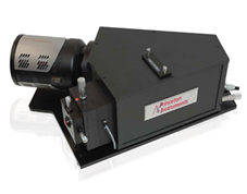
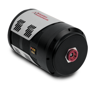
In their measurements the researchers use a setup scanning a fiber optic probe over tissue samples. The probe design contains fibers for FLIM and Raman excitation and signal collection as well as filters required in Raman spectroscopy for clean laser excitation and suppression of elastic scattered light. As FLIM is the faster of the two techniques, it is used to scan the surface and identify targets (detecting changes in fluorescent lifetime) to be investigated using Raman spectroscopy by excitation with a 785nm multimode laser. The Raman signals are obtained using a LS-785 spectrograph and PIXIS camera.
Advanced numerical techniques (extended multiplicative signal correction EMSC, partial least-squares linear discriminant analysis PLS-LDA) are used for systematic background subtraction and data analysis. The changes in the Raman spectra correctly identify cross-linked from non-cross-linked tissue and provide the researchers high biochemical sensitivity to identify calcification, while FLIM is shown to better detect the degree of cross linking in the collagen matrix.
The authors conclude that “FLIM and Raman imaging has a strong potential to address some of the pressing needs for nondestructive and label-free monitoring of tissue engineering processes and their applications in regenerative medicine.” Combining both measurement technique leads to a higher understanding of the biochemical and structural processes of BP, helping in studying the performance of the tissue over time.
Researchers around Suotang Jia in China investigate the behavior of single quantum dots in solvents designed to suppress photobleaching and blinking.
Quantum dots are now widely used in applications such as light sources, improving the efficiency of photovoltaic cells or fluorescent markers in biomedical applications. Blinking and photobleaching are significant challenges to overcome to use the full potential of quantum dots in these applications. The researchers use microscopic widefield imaging and confocal microscopy to observe the emission of single quantum dots. Sensitive EMCCD cameras are well suited to measure fast sequences of images of these single molecule emitters.
Combustion processes in real world applications, such as motors and engines, typically involve turbulent flow patterns and complex reaction chemistries in high and lower temperature flames. The lab of Prof. Yiguang Ju from Princeton University just reported on experiments measuring the behavior of flames interacting with a vortex (simulating turbulence). One of the research goals of the Ju lab is the exploration and development of new knowledge and techniques to advance new and cleaner combustion techniques. Moreover, the lab has been experts in creating cool diffusion flames and investigating turbulent combustion processes.
The lab created double – cool and hot – flame and observed as the flame interacts with a flow vortex. The researchers use a planar laser induced fluorescence technique (PLIF). A pulsed laser beam is expanded into a two-dimensional sheet of light that is send through the flame. The fluorescence signal is measured with a gated PI-MAX4 ICCD camera. PLIF allows to target specific molecule species and results in a two-dimensional fluorescence image. Besides high temporal and spatial resolution PLIF allows researchers to measure molecule concentrations, distributions and velocities as well.
Rose Padilla and their team/collaborators report on experiments where they measure methane/air combustion flames mixed with water. The goal is to understand the effects of having water on the fuel side where in real world situations the water can be introduced arbitrarily or occurs naturally as in methane hydrates.
The researchers measure the PLIF of OH radicals to characterize the flame reaction zone using a PI-MAX4 ICCD camera. The same camera is used for chemiluminescence CH molecules as well.
In the field of Plasma physics ICCD cameras are often used for fast time resolved imaging due to their ability to take quick snapshots of dynamic reactions.
In this paper researchers use a PI-MAX camera for time resolved imaging of plasmas from different electrode structures. Their goal is to characterize different plasma producing electrodes for applications in life science and biomedicine.
Researchers from RWTH Aachen in Germany are trying to understand how soot forms in combustion processes. Soot is typically an unwanted by product of the combustion process and a major contributor to environmental and health hazards associated with it. As is common practice in combustion experiments the researchers use intensified cameras to achieve precise time resolution as well as distinguishing signals from the bright flame background when they are excited by very short laser pulses.
For example, monitoring fluorescence from two, narrow band spectral lines using custom filters allows for measurement of the local gas temperature. The distribution of OH was measured using narrow band filters in the UV around 310 nm using planar laser induced fluorescence.
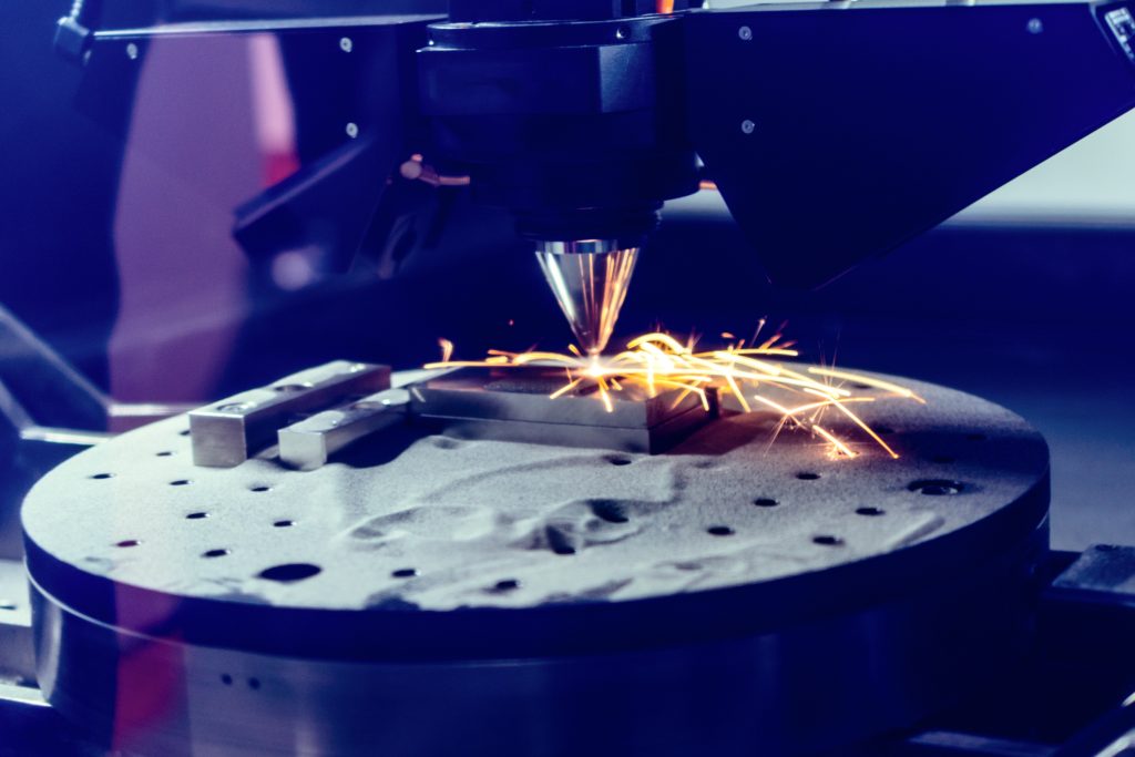
Laser based manufacturing processes like cutting, welding and recently additive manufacturing are key techniques in the manufacturing world with a multi-billion-dollar market. However, to further optimize and advance these methods a detailed understanding of the light material interactions must be obtained theoretically and experimentally. Ideally one can observe the interaction process in real time, however the time scale of the interaction process is in the nanosecond range, so imaging is challenging.
Experiments at the Dynamic Compression Sector of the Advanced Photon Lightsource of Argonne National Lab use a total of 4 ICCD cameras to accomplish this task. The cameras are operated in a double image feature mode that allows to take 2 consecutive images with as low as 450ns time between images. The X-Ray radiation monitoring the materials is converted by a scintillator into visible light that and guided through beam splitters that are aligned such that each camera is observing the identical section of the sample. By precise, correlated triggering of the cameras a total sequence of 8 images with less than 100ns time resolution can be acquired to study the ultrafast dynamics of the laser interaction process.