emICCD Cameras Facilitate Use of Trapped Ions for Quantum ResearchApplication Notes

Introduction

Laser-cooled ions in linear Paul traps are quantum systems with remarkable properties. Trapped ions offer an unprecedented degree of preparation and control of their parameters, can be cooled to the ground state, and can be coupled to engineered reservoirs. For these reasons, they have played a prominent role in the experimental study of quantum computation and information processing applications. They are also invaluable tools for the investigation of quantum thermodynamics.1


Today, researchers in many fields use trapped ions to perform experiments that enable the investigation of quantum phenomena. The list of applications employing trapped ions is growing quickly and this type of work likely holds the key to major advances in such diverse areas as quantum computing, quantum cryptography, and light-based telecommunications. The present application note will focus on the research being conducted jointly by scientists from two universities in Germany (Mainz, Augsburg). This group of scientists recently proposed an experimental scheme to realize a nano-heat engine with a single ion.1

Quantum Heat Engines

The fundamental question that the aforementioned group of scientists seeks to address is whether commonly realized macro-heat engines like those utilized by automobiles, which convert thermal energy into mechanical work, can be scaled down to the single-particle level while retaining the same working principles. The scientists have analyzed an experimental scheme for a nano-heat engine using a single laser-cooled ion as working gas.

In experiments performed at the Institut für Quantenphysik, Universität Mainz, the team has implemented an Otto cycle, the basis of the four-stroke car engine, by confining the cold ion in a linear Paul trap with a tapered geometry (see Figure 1) and coupling it to a pair of engineered reservoirs.

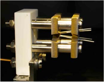
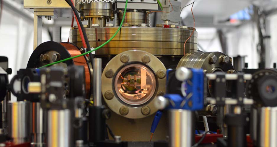
Figure 1. Left: A linear Paul trap for the singleion heat engine. Its tapered geometry allows the coupling of radial states to the axial oscillation.2 Right: Experiment setup showing the Paul trap in a vacuum chamber. Photos courtesy of Prof. Dr. Ferdinand Schmidt-Kaler, Dr. Kilian Singer, Johannes Rossnagel, and Georg Jacob (Institut für Quantenphysik, Universität Mainz)

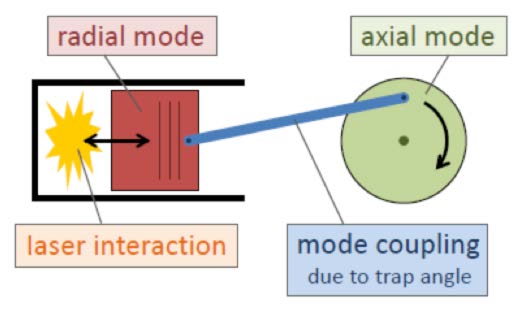
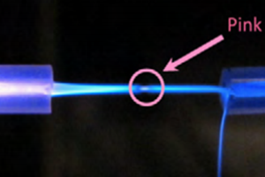
The two heat baths alternately heat and cool the radial thermal state of the ion. The change of radial temperatures is converted into axial movement — and thus into usable work. By repeating this cycle resonantly with the axial eigenfrequency at 100 kHz, large coherent amplitudes along the trap axis are accumulated.2 A classic analog is presented in Figure 2.

Figure 2. A classic piston/flywheel analog.2 Diagram courtesy of Prof. Dr. Ferdinand Schmidt-Kaler, Dr. Kilian
Singer, Johannes Rossnagel, and Georg Jacob (Institut für Quantenphysik, Universität Mainz).

The German scientists have analytically determined the quantum efficiency of the single-ion heat engine at maximum power in various regimes. They have also performed Monte Carlo simulations of the engine that not only demonstrate its feasibility but its ability to operate at maximum efficiency of 30% under realistic conditions.1


Although micro-heat engines have been constructed employing various types of systems, a quantum heat engine had never been built. Encouraged by their numerical simulations and analysis, however, the scientists in Germany have now fabricated a single-atom engine.3

Experiment Imaging

In their experiments, a single calcium ion is trapped in the electromagnetic field of a special Paul trap and cooled via laser to a temperature of 1 mK. To drive the ion in a heat engine cycle, the scientists alternately heat and cool the ion by applying electric noise and laser cooling.


The figure of merit for the experimental demonstration of the single-ion heat engine is the work produced as a function of the temperatures of the heat baths. As the work produced is directly converted into motion in the form of an oscillation at 100 kHz, a precise observation of the latter is crucial.

In order to observe the oscillation of the ion at 100 kHz, the scientists employ an emICCD camera. Exposure times of only 0.5 μsec allow the ultra-high-sensitivity imaging system to resolve the ion motion precisely in both space and time (see Figure 3).


It is the fast gating ability of the emICCD camera that has enabled the scientists to demonstrate the functionality and performance of the single-ion heat engine. This will lead to a further understanding of the thermodynamics of single particles.

Figure 3. Screenshot from a video imaging individual ions, acquired with a Teledyne Princeton Instruments emICCD camera. Courtesy of Prof. Dr. Ferdinand Schmidt-Kaler, Dr. Kilian Singer, Johannes Rossnagel, and Georg Jacob (Institut für Quantenphysik, Universität Mainz)

Enabling Technology

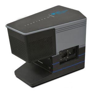
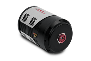
The PI-MAX4:1024EMB emICCD camera (see Figure 4) seamlessly combines the rapid gating capabilities of an image intensifier and the excellent linearity of a back-illuminated, frame-transfer, 1024 x 1024 EMCCD detector in order to deliver quantitative, ultra-high-sensitivity performance for applications executed on nano- and picosecond timescales.

Figure 4. The PI-MAX4:1024EMB from Princeton Instruments gives users the benefits of both an intensified CCD (ICCD) camera and an electron-multiplying CCD (EMCCD) camera.

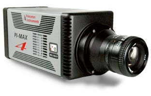
This fiberoptically bonded Princeton Instruments PI-MAX®4 camera system provides <500 psec gate widths using standard fast-gate intensifiers while preserving quantum efficiency. Its integrated SuperSynchro timing generator allows camera users to set gate pulse widths and delays under GUI software control, and significantly reduces the inherent insertion delay (~27 nsec).

Complete control over all PI-MAX4:1024EMB hardware features is simple with the latest version of Princeton Instruments’ LightField® data acquisition software (available as an option). Precision intensifier gating control and gate delays, as well as a host of novel functions for easy capture and export of image data, are provided via the exceptionally intuitive LightField user interface.

The PI-MAX4:1024EMB uses a high-bandwidth (125 MB/sec or 1000 Mbps) GigE data interface to afford camera users real-time image transmission. This interface supports remote operation from more than 50 meters away.

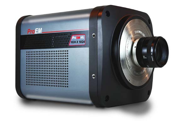
Alternatively, research that does not require gating can benefit from advanced high-speed EMCCD technology such as that implemented in Princeton Instruments’ ProEM-HS cameras (see Figure 5). The ProEM® “HS” series offers a special kinetics readout mode that increases temporal resolution by illuminating a small portion of the sensor and then capturing and shifting a series of sub-frames in microseconds. These high-sensitivity, back-illuminated EMCCD cameras are engineered to keep up with the latest high-repetition-rate lasers.

Figure 5. ProEM-HS cameras from Princeton Instruments offer the high sensitivity of back-illuminated, electron-multiplying CCDs and the speed of a special kinetics readout mode.

Future Trends

As myriad technologies, devices, and systems march inexorably toward the ultimate limit of single quantum particles, experimental studies will become more challenging and complex. Accordingly, next-generation scientific cameras must be able to provide the outstanding sensitivity and speed demanded by such investigations. The PI-MAX4:1024EMB and ProEM-HS cameras mentioned herein represent the leading edge of high-performance quantitative imaging capabilities for research utilizing trapped ions

References

  1. Abah O., Rossnagel J., Jacob G., Deffner S., Schmidt-Kaler F., Singer K., and Lutz E. Single-ion heat engine at maximum power. Phys. Rev. Lett. 109, 203006 (2012). DOI: 10.1103/PhysRevLett.109.203006
  2. https://www.quantenbit.physik.uni-mainz.de/index.wt/index.wt/#/research/thermo (accessed online June 2015)
  3. Rossnagel J., Abah O., Schmidt-Kaler F., Singer K., and Lutz E. Nanoscale heat engine beyond the Carnot limit. Phys. Rev. Lett. 112, 030602 (2014). DOI: 10.1103/ PhysRevLett.112.030602

Application CTA Section

Ultra-Low-Light Imaging in Quantum Research

Find out about the different types of camera technologies that can be used for quantum research, their advantages and their limitations.

emICCD Cameras for Diamond Quantum Dynamics Research

Discover how researchers from Canada used an emICCD camera to image the quantum state of NV centers in diamond.

<500 Picosecond Gating for Atmospheric Pressure Plasma Jets

An application note providing an overview of the experimental setups for atmospheric pressure plasma jets alongside relevant imaging technology.

Exploring the Physics of 2D Magnets with Sensitive ImagingResearch Stories

Jie Shan

arXiv

Imaging and control of critical spin fluctuations in two-dimensional magnets

Introduction

Researchers at Cornell University have imaged light scattered from a two-dimensional, ferromagnetic material using the ProEM-512 EMCCD camera. The researchers around Prof. Jie Shan from Cornell University (USA) want to better understand the physics of magnetic fluctuations in this material and how to control them which could lead to the realization of magnet/spin-controlled devices in the future.


By using magnetic circular dichroism (MCD) they are able to achieve fast and sensitive imaging. MCD works similarly to circular dichroism but in the presence of a magnetic field. Left/Right-handed circular polarized light is scattered by the oriented atomic spins in the sample and using a setup of well aligned polarizers, regions of different spin orientations can be imaged with very high contrast and sub micrometer spatial resolution.


The samples are single atomic layers of a material called CrBr3 that is encapsuled in thin atomic layers of hexagonal Boron nitride (this is called a van der Waals heterostructure made of two-dimensional materials). Sensitive imaging (10ms exposure time) is aided by the large magnetic moment of this material and helps taking image sequences with changing magnetic field

Further Information

Imaging Diffusion Clouds of Charges in PerovskitesResearch Stories

Rashid Zia

ACS Photonics

Direct Characterization of Carrier Diffusion in Halide-Perovskite Thin Films Using Transient Photoluminescence Imaging

Introduction

As energy production is continuously shifted to renewable energy sources, perovskites are heavily researched for their applications in solar cells promising higher efficiencies than current devices based on silicon. Current research on perovskite-based photovoltaics tries to overcome the short lifetime of devices made of this material as well as understand the detailed physics during the energy conversion process.

Researchers associated with Rashid Zia from Brown University (USA) describe in a recent publication in ACS Photonics that understanding these intrinsic properties helps improve design, engineering and fabrication of perovskite-based devices as well as guide further developments in this area.

Furthermore, the absolute limit of efficiency achievable with perovskite-photovoltaics is not yet known. The transport of charge carriers is a main influence of device performance. To quantify the behavior of transport in perovskites the team around Prof. Zia has developed a time-gated photoluminescence imaging technique with sub nanosecond time resolution. For detection their setup uses a PI-MAX4 emICCD with gate width down to 500ps.

To acquire the weak photoluminescence signal the camera operates by accumulating signal from several hundreds of thousands of photoluminescence decays acquired with 10kHz repetition rate. By changing the delay between excitation laser pulse and gate opening the diffusion of charge carriers can be directly imaged on the ns level. The technique is compatible with low signal emission from the rapidly diffusing charge carriers unlike transient absorption measurements. Also, the technique speeds up data acquisition by observing a large sample area unlike point scanning methods based on fast photodetectors such as avalanche photodiodes.

Further Information

Comparing Human and Emperor Penguin EnzymesResearch Stories

John C. Hackett

Journal of Inorganic Biochemistry

Biophysical characterization of Aptenodytes forsteri cytochrome P450 aromatase

Biological systems and molecules can be used as model systems for biochemical and physiological processes in humans if they are genetically/evolutionary related to similar molecules. The research team of John Hackett investigated the similarities of enzymes of emperor penguins to human enzymes using absorbance and Raman spectroscopy.

They perform resonance Raman spectroscopy, using a SpectraPro spectrometer and a liquid nitrogen cooled PyLoN CCD camera, with excitation in the UV (406nm) close to electron transition energies of the enzyme. The Raman spectra are sensitive to small molecular and structural changes of the molecules. High resolution spectroscopy with a sensitive detector is used to acquire spectra.

Further Information

Enhancing SWCNT Properties Through AlignmentResearch Stories

Bill Rice

arXiv

Global alignment of solution-based single-wall carbon nanotube films via machine-vision controlled filtration

Introduction

Bill Rice’s group at the University of Wyoming is investigating the physical properties of unique and interesting materials. One of their research projects involves single walled carbon nanotubes (SWCNTs). These small, 1D crystals of rolled up graphene sheets come with a wide variety of physical properties (depending on the roll direction) and research has made great advances in purifying unique nanotubes which would be ideal for real world applications. However, a common nanotube characteristic is that their behavior is very different along their axis showing enhanced physical effects in comparison to the perpendicular direction showing reduced physical effects.

The group is building films of highly aligned nanotubes for optimal amplification of their physical properties and they are researching for robust, fast methods to monitor the quality of alignment. As is often the case for carbon materials, Raman spectroscopy plays a crucial role in determining the orientation of crystals by using polarized Raman signals which are strong if the polarization direction is parallel to the nanotube axis. The results of polarized Raman measurements build the reference to characterize other methods for process control and monitoring. The Raman spectroscopy setup is realized using an Isoplane320 spectrograph and liquid nitrogen cooled PyLoN camera.

Further Information

Portable System for Fusion Prototype CharacterizationResearch Stories

Theodore Biewer

Oak Ridge National Laboratory

ORNL team builds portable diagnostic for fusion experiments from off-the-shelf items

Oak Ridge National Labs (USA) have developed a portable plasma measurement system, built by Theodore Biewer and his team. Plasmas are a key technology for potential energy production (fusion research), propulsion systems and material processing.

The system will be taken to different experiment sites to measure plasma properties specifically the density of free electrons and temperature using Thomson scattering (elastic light scattering by charged particles).

The setup uses aberration corrected spectrographs (Isoplane-320) and intensified CCD cameras (PI-MAX4) for detection. Low aberration spectroscopy allows for simultaneous detection of multiple spatial locations in the plasma while the ICCD camera suppresses the background radiation of the hot ion gas.

Further Information

UV Resonance Raman for DNA Structure StabilizationResearch Stories

Bottari C. et al.

SPIE

Conformational stability of DNA in hydrated ionic liquid by synchrotron-based UV resonance raman

Introduction

A group of Physicists and Chemists from Italy are using CERIC-ERIC Consortium for studying conformational stability of DNA. Their research investigates ionic liquids as next generation solvents to stabilize the double-helix structure of DNA.

They use synchrotron-based UV Resonance Raman (UVRR) spectroscopy with the aim of clarifying the role played by concentration of ionic liquids (IL) in stabilizing the natural conformation of DNA. UVRR spectroscopy has been powerful tool to researchers in the field of protein structural analysis. This technique is able to selectively enhance the vibrations mainly localized on nucleotide rings, when in resonance conditions, thereby minimizing interference associated with phosphates and sugar backbone.

UVRR measurements allowed them to enhance specific vibrational signals associated to nitrogenous bases of DNA, through an appropriate tuning of the excitation wavelength which was possible using our tunable band pass filter Trivista 557 spectrograph. The radiation power on the sample was ~20 W. Any possible photo-damage effect due to a prolonged exposure of the sample to UV radiation was avoided by continuously spinning the sample cell during measurements.

Further Information

HyNASDs As ASD Alternative For Enhanced Release of BCS Class II DrugsResearch Stories

Ecevit Bilgili

European Journal of Pharmaceutics and Biopharmaceutics

Hybrid nanocrystal–amorphous solid dispersions (HyNASDs) as alternative to ASDs for enhanced release of BCS Class II drugs

Hybrid nanocrystal-amorphous solid dispersion (HyNASDs) has been utilized, via the FERGIE spectrometer (a previous version of the IsoPlane 81), to study the enhancement of bioavailbaility of poorly soluble drugs.

Researchers from the group of Ecevit Bilgili at New Jersey Institute of Technology, USA, determined that using a hybrid mixture of nanocrystal along with an amorphous dispersion in the dissolution media enhances the release of BCS class II drugs as compared to conventional amorphous solid dispersions. This was demonstrated by measuring the percentage of griseofulvin that supersaturated in dissolution.

The group has studied solid state drug-polymer interactions utilizing Raman spectroscopy in combination with XRPD (x-ray powder diffraction) and DSC (differential scanning calorimetry) measurements. Their Raman measurements used a 785nm, 500-mW external diode laser along with the FERGIE spectrometer, with data acquisition at ~15 sec per spectrum.

The group is actively involved in fundamental understanding of the mechanisms behind the transformation of drug nanoparticles via top-down and bottom-up approaches. Raman spectroscopy has helped them in understanding the drug-polymer interaction which was key to understanding spray dried sample compositions in comparison to samples from nanosuspension feed

Further Information

Raman Spectroscopy for Skin Wound EvaluationResearch Stories

Quan Liu

The Journal of Physical Chemistry Letters

Depth-Sensitive Raman Spectroscopy for Skin Wound Evaluation in Rodents

Introduction

Researchers at Nanyang Technological University in collaboration with others demonstrate design/optimization of a new snapshot depth sensitive Raman instrument for skin sample measurements. Their setup uses the Acton LS785 Spectrograph and PIXIS camera for Raman spectroscopy. Their depth sensitive measurement performance is characterized in both an ex vivo porcine skin with a gradient of layer thickness and an ex vivo rat skin sample with surface wounds. The exposure time for their experiment was well below the permissible exposure as per American national standards.

By using a signal collection fiber bundle, the researchers were able to adjust for variable depth profiles on the sample. They demonstrate simultaneous data acquisition at five different target depths of the sample. This correlates to very good efficiency for an in vivo scenario. PLS-LDA analysis created classifiers between the wound edge and healthy skin based on the acquired spectra. The instrument can discriminate between the wound edge and healthy skin with good accuracy which paves the way towards in vivo preclinical studies of rat skin wounds.

Acton LS
PIXIS photo

Further Information

Microspectroscopy for Characterizing Nanostructure CouplingResearch Stories

Jing Zhao

The Journal of Physical Chemistry Letters

Colloidal Assembly of Au-Quantum Dot-Au Sandwiched Nanostructures with Strong Plasmon-Exciton Coupling

Introduction

This summary is from an article by researchers around Prof. Jing Zhao from the University of Connecticut. Her group is investigating interactions between quantum dots and metallic nanoparticles that are fundamental for nanostructure applications for light emission, energy production and optical sensing.

Optical investigation and spectroscopy are essential tools for characterizing structures and particles on the nanometer scale and are typically coupled with microscopic measurement to ensure precise localization of the structure of interest. The spectrum of light scattered and emitted by nanoscale systems depends predominately on their structure and environment. By comparison of the measured spectra with models and simulations, the internal physics of the structures is revealed.

Prof. Zhaos groups setup is built around an inverted microscopy (Nikon Ti-U) that is connected to an imaging spectrograph (Isoplane SCT 320) with an imaging CCD detector (PIXIS 1024). The Isoplane is an ideal system for use in microscopy applications, using large area detectors, as it is not limited by image aberrations that reduce the spectral quality, resolution and sensitivity.

The coupling between the QDs and metallic nanoparticles measures the transmission of a broadband lamp alongside the fluorescence when excited with a short-pulsed laser system. Using this measurement system, the scattering signal and background can be acquired at the same time by measuring 2 adjacent sections of the sample simultaneously on the detector.

The measurements allowed the team of Prof. Zhao to characterize strong coupling of their nanostructures demonstrating the ability of the sample production methods to precisely arrange the nanoparticles.

Further Information