X-ray Imaging
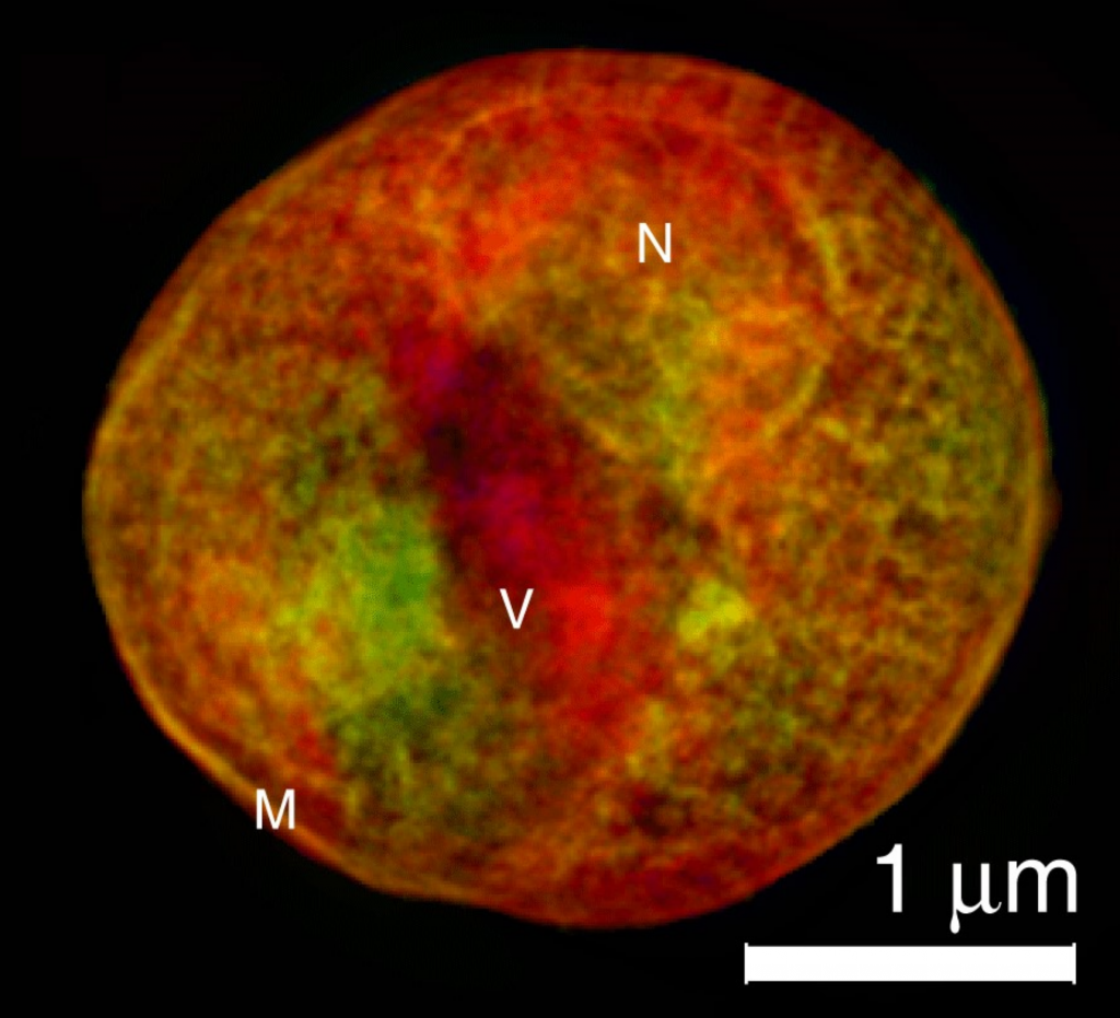
Due to their short wavelengths, x-rays can be used to image samples on the nanoscale. There are multiple different types of x-ray imaging, such as x-ray tomography, or coherent diffraction imaging. Imaging can be combined with microscopy or spectroscopy to find out further information about a sample, such as chemical composition.
X-ray tomography enables 3D reconstruction of internal structures of real objects non-destructively, all while maintaining a high spatial resolution. By detecting either the attenuation or the phase shift of the transmitted beam at various angles, 2D slices of the microstructure of a material can be reconstructed. These 2D slices can be stacked together to produce a 3D image of the material.
Coherent diffraction imaging (CDI) is a lensless technique which reconstructs an image from the diffraction patterns of a sample. Reconstruction of the image is achieved via an iterative feedback algorithm which retrieves the lost phase information. In this way the lens of an instrument is replaced by the algorithm. CDI is able to provide 2D and 3D information about a material, providing high spatiotemporal resolution for probing dynamic phenomena.
Direct X-ray Detection
SOPHIA XO
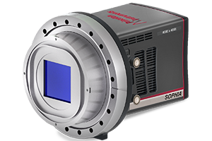
X-ray imaging is sample dependent, so relies on high quantum efficiency, and experimental flexibility. The SOPHIA-XO offers excellent sensitivity, with a peak quantum efficiency >95%. The SOPHIA-XO also has multiple port readouts and high frame rates, minimizing overall experimental time, essential for imaging of large samples.
To ensure all x-ray signal is captured, the SOPHIA-XO comes with both 2k x 2k and 4k x 4k sensor options. With low read noise and deep cooling to minimize dark current noise, the SOPHIA-XO allows for the detection of the faintest of x-ray signals.
PI-MTE3
The PI-MTE3 can be used fully in-vacuum, including the cooling electrical cables, giving exceptional reliability for flexible camera placement within a vacuum chamber. With large sensor formats, of up to 4k x 4k resolution, the PI-MTE3 is able to capture fine details within x-ray imaging.
The PI-MTE3 is liquid cooled, resulting in low dark current over long integration times, for detection of faint x-ray signals. With 7-10x higher resolution than previous PI-MTE generation, the PI-MTE3 achieves 3 full fps at 2k x 2k resolution, reducing overall readout times in comparison to previous generations.

PIXIS XO
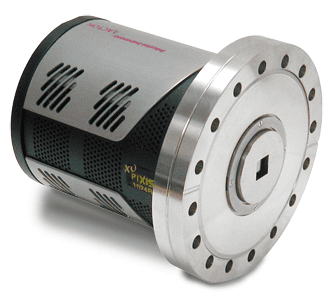
X-ray imaging relies on high resolution and sensitivity for accurate image reconstruction. The PIXIS XO offers excellent resolution and sensitivity for soft x-rays, directly detecting over a wide range of energies.
The PIXIS XO comes with multiple CCD sensor options, all without antireflective coating, for ultimate experimental optimization. The ultra-high-vacuum-seal design further enhances the flexibility of the PIXIS XO, with software-selectable gains and readout speeds.
Indirect X-ray Detection
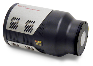
PIXIS XF
The PIXIS XF uses fiberoptic coupling to preserve the highest spatial resolution, imaging indirect x-rays in the < 3 keV to >20 keV energy range – ideal for Micro-CT.
The unique mechanical design allows for outstanding flexibility. Specialized, removable phosphor screens allow for optimization of the system, with the PIXIS XF providing multiple sensor options, including front- or back-illuminated CCDs for varying experimental design.
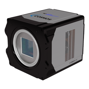
COSMOS
COSMOS is a large format CMOS camera, with sensor sizes ranging from 3k x 3k to 8k x 8k in resolution. COSMOS offers superior sensitivity, < 0.7 e- read noise, and fast frame rates ideal for imaging large sample areas while maintaining high spatial information and resolution.
With both a wide dynamic range, and unsurpassed linearity, COSMOS is able to provide exceptional quantitative precision across all measurements, even for faint signals. Deep cooling of COSMOS reduces the thermal dark current noise generated, further minimizing any noise artifacts.
