Dr. András Deák
Centre for Energy Research, Eötvös Loránd Research Network
Background
Dr. András Deák’s research focusses on understanding the physics behind how molecules interact and attach to nanoparticle surfaces. Many applications rely on introduced molecules attaching to the surface of a nanoparticle in a predetermined way. However, if a nanoparticle already has molecules attached to its surface, complete molecular exchange cannot be guaranteed. Single nanoparticle scattering experiments can provide insight into the extent of molecular exchange, with peak shift and broadening correlating to an increase in molecular attachment. The advantage of this approach is that it is not affected by the inhomgeneous broadening inherently present during the commonly used bulk, ensemble measurements.
Another aspect of András Deák’s research focusses on self-assembly, that is, how nanoparticles interact with each other in solution. Nanoparticles are fabricated and surface modified so that they will attach to each other in a predefined way. However, this attachment is often characterized via scanning electron microscopy (SEM), a process which removes the nanoparticles from their liquid environment. As the environment the nanoparticles are being characterized in has changed, it is uncertain whether this is truly how the nanoparticles interact within the liquid medium.

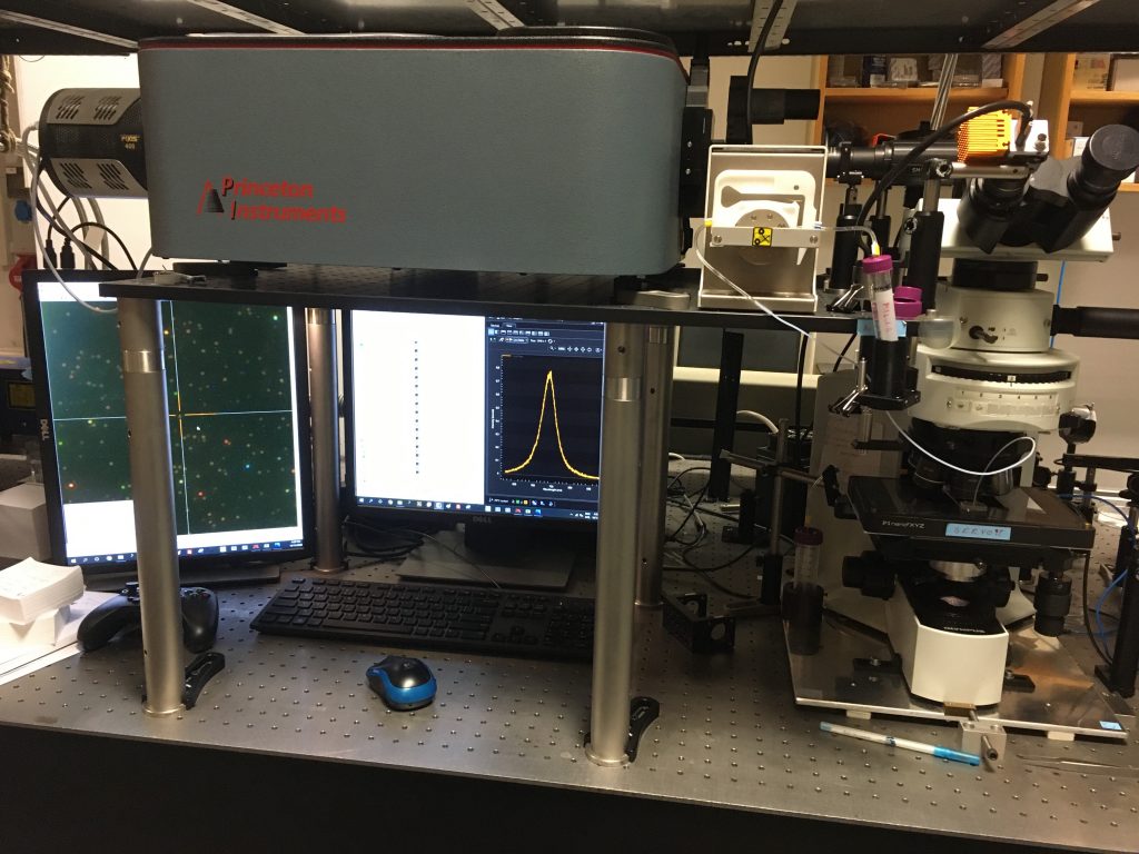
Figure 1 : An image of the custom built microspectroscopy system, comprised of the IsoPlane and PIXIS CCD camera attached to an optical microscope. The spectrum shows the redshift of the longitudinal surface plasmon resonance peak of an individual gold nanorod upon reacting with 1 mM cysteamine.
Challenge
In order to overcome both inhomogeneous scattering and environmental changes caused by SEM characterization, András Deák has developed a custom built microspectroscopy system. This system is set up to analyze the scattering spectrum of individual nanoparticles via dark field illumination.
When looking at molecular exchange, the researchers place nanoparticles with molecules attached to the surface into a flow cell under an optical microscope. They then introduce new molecules into the system and monitor how the scattering spectrum of the individual nanoparticle changes over time. The width of the scattering peak correlates to the number of molecules that have attached to the surface, so can be used to determine the level of molecular exchange.
This method can also be used to determine how nanoparticles interact within liquid on the single nanoparticle scale. By using optical spectroscopy, András Deák’s researchers can look at the surface details of individual nanoparticles without the need to remove them from the liquid medium. Any changes in the spectrum of these nanoparticles indicates that the nanoparticles have interacted with each other.
As the IsoPlane is aberration corrected, we can have highly reliable intensity values over the whole range of the CCD
Solution
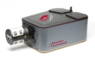
The custom microspectroscopy system is comprised of an IsoPlane and PIXIS CCD camera attached to an optical microscope, onto which a flow cell with the nanoparticles is placed. The high flexibility of this system allows for the integration of the different components via LabVIEW. This allows for full control over the IsoPlane, PIXIS, piezo stages and rotational stages from one program.
The IsoPlane is ideal for nanoparticle microspectroscopy as it is aberration corrected, producing an accurate spectrum for each nanoparticle without any additional spectral broadening. This also prevents the spectrum from spreading on the CCD, providing highly reliable intensity values that cover the whole wavelength range captured by the PIXIS. The system also uses an IntelliCal wavelength and intensity calibration lamp, allowing researchers to take very reliable measurements rapidly. This allows the researchers to evaluate the physics of nanoparticle surfaces on the single nanoparticle scale with high accuracy very quickly.

